


Say goodbye to re-occuring calf injuries
Persistent calf injuries are a common complaint amongst runners and sports people. Second only to the knee, the lower leg accounts for approximately one-third of running injuries in long-distance runners (1, 2). There are numerous causes of lower leg pain, with diagnoses varying in frequency and severity. The most common causes include; calf strains, medial tibial stress syndrome (MTSS), chronic exertional compartment syndrome (CECS), stress fracture, nerve entrapment, and popliteal artery entrapment syndrome (PAES) (3). These lower leg injuries can often be a persistent cause of frustration for any athlete if not promptly diagnosed and managed. If you have had calf troubles, whether it be pain, cramping or tightness it is time to get it sorted.
What is the diagnosis?
A common mantra around the practice is “diagnosis determines prognosis.” There is no overstating the importance of the vital first step – diagnosis, alongside a thorough assessment will determine best management and give realistic time frames for return to running, sport and full function.
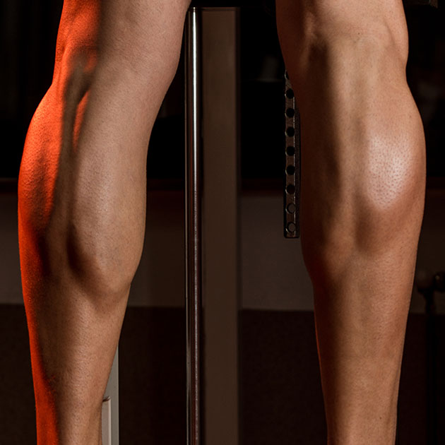
A common mantra around the practice is “diagnosis determines prognosis. #performbetter @pogophysio Share on XThe ‘‘calf muscle’’ consists of three separate muscles; the gastrocnemius, soleus, and plantaris which unite to form the Achilles tendon. Differentiating strains in the gastrocnemius and soleus is particularly important for an accurate prognosis, appropriate rehabilitation (4). The gastrocnemius has a higher portion of fast-twitch muscle fibres (type 2) and crosses two joints (the knee and ankle). It is classically injured by quickly and powerfully extending the knee with a dorsiflexed (bent ankle), for example trying to accelerate quickly from a stationary start into a run. The plantaris is a small accessory muscle which also crosses the knee and ankle. It is difficult to isolate from gastrocnemius injuries without imaging and is rarely injured in isolation (4). The soleus crosses only the ankle and is largely comprised of type one slow twitch muscle fibers. Soleus strains also tend to be less dramatic in clinical presentation and more subacute when compared to injuries of the gastrocnemius. The classic presentation is of lower calf tightness, soreness and stiffness aggravated by walking and running, particularly of a longer duration. Additional details from your history and clinical assessment of strength, mobility and palpation will help in determining diagnosis.
Calf strains can also present similar to other forms of lower limb pain. Some common differential diagnosis for calf injuries can include:
- Medial Tibial Stress Syndrome – often the true diagnosis behind ‘shin splints,’ it involves changes to the peristeoum (bone surface) of the lower inside border of the tibia.
- Stress fractures – following on from a stress reaction the next step in the bony stress continuum is a stress fracture. These can occur along the medial or posterior border of the tibia and can occur following spikes in lower limb loading.
- Chronic Exertional Compartment Syndrome – CECS is characterised by recurrent, often bilateral severe muscle compartment pain that occurs with exercise and subsides with rest. Often there are no other clinically significant signs such as loss of movement or strength decreases (5). Often conservative treatment is unsuccessful and fasciotomy (surgical resection of the fascia surrounding the muscle belly) is treatment of choice.
- Nerve Entrapment – The common peroneal, superficial peroneal, and saphenous nerves are most commonly at risk for entrapment. These are quite rare and are often preceded by trauma. Burning pain, sensory and/or motor (strength) changes may occur (4).
- Popliteal Artery Entrapment Syndrome – PAES is an important and often over-looked differential diagnosis in the assessment of exertional leg pain. PAES is defined as a group of conditions in which compression of the popliteal artery, popliteal vein, and/or tibial nerve, in the popliteal fossa by surrounding musculoskeletal structures occurs to a degree sufficient to cause vascular and neurogenic symptoms (6).
Determining Prognosis
Recovery time frames are dependent on degree of calf injury. Healing timeframes will be influenced by grade of muscle tear; commonly referred to as either a minor partial tear (Grade 1), moderate partial tear (Grade 2) or complete muscle tear/tendon avulsion (Grade 3) (4). These classifications are based off clinical signs and symptoms, combined with diagnostic imaging (US or MRI). Recovery time frames will also be influenced by injury involvement to the musculotendinous junction, age, past history of injury and need to address contributing factors.
What are the risk factors?
Athletes, coaches, trainers and physiotherapists alike are always looking to reduce injury risk. Predictors of calf injury are tough to find in the literature. However a study by Van Middelkoop et al. found that a lower extremity injury in the previous 12 months was a risk factor for a knee injury, and that an injury at another location (hip, groin, thigh, knee, ankle, or/and foot) was a risk factor for calf injury (8). In addition to previous injury (calf or other), other factors that may increase risk of injury include; older age, calf muscle weakness, increases in training loading (find your optimal training load) or altered loading mechanics (see below).
Management
Acute management aligns to the management of muscle injuries. Over the first 3–5 days, muscle rest by limiting stretch and contraction, cryotherapy (ice), compressive wrap or tape, and elevation of the leg are generally recommended (4). Depending on degree of injury and pain a heel wedge or crutches may be useful. Heat, massage and NSAIDS are often avoided in early stage of management. Over the coming days to weeks strengthening and stretching are commenced. Return to running or activity is often begun after full range and strength is returned. Strengthening should be targeted to both gastrocnemius and soleus (by altering degree of knee extension), with additional focus on the injured muscle. It is then important to consider the demands of your chosen sport, do you need to work on power, endurance, multi-directional stability or particular surfaces/terrain.
How well do you know your running body? (compensations/biomechanics)
For many people a calf injury can be a simple as the above, you strain the muscle, rest then stretch and strengthen, then you get back to normal activity. In other cases the calf pain returns, it is re-injured or continues to niggle feeling tight or sore. This is where knowing your body’s biomechanics and running patterns become important. Forward propulsion can occur in three ways, you fall forward, you get pushed forward, or you push yourself forward. How do you do this? Your posterior chain – glutes (glute max with some help from glut med/min), hamstrings and calf (gastrocnemius, soleus). Changes or imbalances from left to right side can lead to one calf working harder, fatiguing quicker, tightening and resulting soreness or injury. Looking biomechanically some reasons may include:
You strain the muscle, rest then stretch and strengthen, then you get back to normal activity #performbetter @pogophysio Share on X- Tight hip flexors – reducing hip extension, calf is working harder for propulsion
- Glut max weakness – as above, tight at the front weak at the back, means the calf is compensating.
- Hamstring Weakness – again, propulsion is hamstrings, glutes and calf – need all three to help
- Excessive ‘bopping’ – increased vertical oscillation (up and down) – pushing ‘up’ is more challenging than pushing forward (try a single leg calf raise standing upright, then at a 30 degree lean and tell me the latter it isn’t easier?)
- Early heel rise – it may be a loss of ankle (dorsiflexion) range, midfoot stiffness, early engagement of the gastrocnemius/soleus, overactive long toe flexors, midfoot pathology forcing early heel rise to avoid pain, reduced 1st MCP joint (big toe) range – numerous strategies can cause this early heel rise, forcing more of a vertical ‘toe off’ phase, reducing the propulsive help of the glutes and again increasing calf loads.
- Anterior compartment weakness – the calf (posterior compartment) and the anterior compartment of the leg are a pair and a balance of anterior – posterior strength is needed.
So if you’re managing a first time calf injury, or an eighth make sure you know it’s your calf, strengthen it and match it to the demands of the sport. Importantly consider what contributed to it and look closer at your biomechanics, so you can say goodbye to future calf troubles.
Lewis Craig (APAM)
POGO Physiotherapist
Masters of Physiotherapy

References
- MacIntyre JG, Taunton JE, Clement DB, et al. Running Injuries: a clinical study of 4,173 cases. Clin J Sport Med. 1991;1:81-87
- van Gent R, Siem D, van Middelkoop M, van Os A, Bierma-Zeinstra S, et al. (2007) Incidence and determinants of lower extremity running injuries in long distance runners: A systematic review. Br J Sports Med 41: 469–480. PMID: 17473005
- Brewer, R. B., & Gregory, A. J. (2012). Chronic Lower Leg Pain in Athletes A Guide for the Differential Diagnosis, Evaluation, and Treatment. Sports Health: A Multidisciplinary Approach, 4(2), 121-127.
- Dixon, J. B. (2009). Gastrocnemius vs. soleus strain: how to differentiate and deal with calf muscle injuries. Current reviews in musculoskeletal medicine, 2(2), 74-77.
- Shah, S. N., Miller, B. S., & Kuhn, J. E. (2004). Chronic exertional compartment syndrome. American journal of orthopedics (Belle Mead, NJ), 33(7), 335-341.
- Hislop, M., Kennedy, D., & Dhupelia, S. (2014). Functional popliteal artery entrapment syndrome: A review of the anatomy and pathophysiology. Journal of Sports Medicine & Doping Studies, 2014.
- Van Middelkoop M, Kolkman J, van Ochten J, Bierma-Zeinstra S, Koes BW (2008) Risk factors for lower extremity injuries among male marathon runners. Scand J Med Sci Sports 18: 691–697. doi: 10. 1111/j.1600-0838.2007.00768.x PMID: 182667


Interesting article,
What is the non – surgery treatment for CECS?
I appear to have triggered it running, but could still cycle – plus calves felt firmer than normal.
I intend to trial massage with gentle stretching and try running again.
Thanks
Thankyou for your question Bill. There isn’t much evidence in favour of non-surgical treatment in true CECS. The best reduction in symptoms comes through activity modification (ie as you have done moving from running to cycling as it has lower risk of increasing compartmental pressure) and rest. Other non-surgical options include mobility work (massage, stretching and rolling), footwear modification (including orthotics) and anti-inflammatories.
Lewis Craig
Physiotherapist
My calf strain reoccurs every time I start running
I gave up in the end trying to run so I started cycling instead. I have strengthened my calf’s using weight training over the years but it still reoccurring.
It can even go these days just walking around a golf course. I would really like to run again can anyone help.
I’m 54 year old male
Hi Stephen,
Sorry to read of your ongoing struggles with your calf strain(s).
I suggest determining if the strains are occurring in the gastrcnemius or soleous muscles (via imaging-if you have a fresh injury).
Strength training wise the target is 0.3-0.4x body weight isolated slow calf and soleous raises in a smith rack at the gym-done x2 per week.
Have a listen to Rich Willy from 39mins in this Episode of our podcast talk about how runners need to strength train HERE>>
Keep me posted.
Regards Brad Beer
I am 54 too and have calf issues. I am a competitive sprint triathlete. My problem usually stems from too much hill or speed work with not enough recovery before my long run. I usually don’t feel the strain from the speed workout, but usually mid way through an 8 mile run a couple days after a harder effort. I am right around 6:50 pace for a 5K. And unfortunately even though this pace feels controlled, my calf’s take a beating. I am really liking the ASICS Glideride series. They are definitely helping relieve pain. Also, I use my theragun everyday before and after runs. Lots of eccentric calf raises too. My PT diagnosed my chronic calf pain as an upper soleus issue. This muscle is more slow twitch and absorbs the landing.
Hi Stephen, I came across your comment and yours and my injuries are identical. We’re also the same age. I’m fed up with mine – tried everything in the last five years and nothing works. Did you ever have any success?
I ran competitively from high school through college and then started triathlon in my mid 30’s it was not until I was 40 I experienced my first soleus strain. It feels like a stabbing pain deep in the lower calf. Over time I strained both right and left soleus repeatedly. Recently I went almost 3 years without an episode, then (always without warning) I pulled the right side. Every time I feel it’s getting better it pulls again. In last 12 months the right side (soleus) has strained at least 3 times. I was hoping to do a marathon in spring but feel hopeless now to train and even more helpless to diagnose why this is happening. Help
Hi Kevin,
I suspect that you may have some underlying soleus weakness (possibly gastrocnemius as well). You would benefit from strength assessment (8RM testing) in a gym for standing and seated calf raises. There are benchmarks for both.
Brad
I live in the US, would you recommend a physical therapist to be an assessor for a runner? I also ran in HS, absolutely loved it, unfortunately stopped, and now I’m in my mid 30s. I’ve ran for almost two years and just recently strained my left calf…again (soleus). Prior to successfully being able to run those two years, I was constantly straining my calf until I modified my gait. I was on a roll, but was unable to pick up any speed, run hills, and my training plateaued. I was just happy to be logging miles until I stained my calf. As I mentioned earlier, I had a MRI and it indicated edema of the soleus and disruption of the central intramuscular tendon. I’ve “rested” for approximately a month, walking, standing at work (office job), found this website which really resonates with me. I’m starting to implement the strength training exercises and walking/jogging. I get so worried about my calf during my running that I can feel myself over-listening to my body. I’m just waiting for that sudden jolt in my calf again…it’s debilitating.
Hi Travis,
Thanks for your question. Yes, I would recommend a physical therapist that has a background or keen interest in running related injuries to perform a formal physical and running assessment on you.
There is no reason why you shouldn’t be able to run injury free again provided you address any strength deficits, optimise your running technique and manage training loads appropriately.
My main tips would be to emphasise your plantarflexor capacity- this will include heavy straight and bent knee heel raises. The soleus generates forces up to 8x bodyweight, even running at slower speeds- therefore don’t be afraid of pushing heavy with the weights. Additionally, improving the Achilles stiffness and elastic recoil will help reduce length excursion of the calf musculature eccentrically upon landing. Slowly progress to exercises such as jump rope and pogo jumps as your strength improves.
Continue building up strength of the entire kinetic chain- including hamstrings, quads, hamstrings and glutes to help absorb running loads.
Your physical therapist will be able to identify any strength deficits that may be predisposing you to recurring calf strains, then formulate an appropriate return to run program.
A gradual return to run program combined with consistent strengthening and time is going to be key to your rehab. It is sensible to first restore your mileage then gradually incorporate things such as speed work and hills which are going to be more demanding on your calves.
Here is a link to our Calf Strain Expert Edition podcast featuring Tania Pizzari and Brady Green: https://www.pogophysio.com.au/blog/tania-pizzari-phd-brady-green/
Hope this helps!
Wow, thanks for this helpful information. Sitting with my second medial calf strain that occurred while playing soccer. Was wanting to get to the answer of why this same part of my calf is quitting ( my teammate diagnosed it as soleus by palpating) and I read here what you say about lack of dorsiflexion straining the calf. Years back exactly on that leg I took a hard ball to a foot that was not planted and toes pointing down. Since that strain I have not been able to properly dorsiflex, almost as if the tibia is jammed forward. But what can I do about that?! Thanks!